Learning is
an act of curiosity.

Sparking Pathshala॥ इतः सम्भवति ॥
- Aorta: Largest systemic artery carrying oxygenated blood under high systolic pressure
- Left Atrium: Receives oxygenated blood from 4 pulmonary veins
- Right Atrium: Receives deoxygenated venous blood via Superior & Inferior Vena Cava
- Left Ventricle: Thickest muscular chamber; pumps blood into systemic aorta
- Right Ventricle: Pumps deoxygenated blood through pulmonary artery to lungs
- Bicuspid / Mitral Valve: Two-cusp valve preventing ventricular backflow into left atrium
3D specimen · click a dot to exploreCor
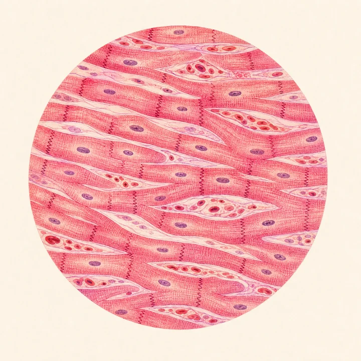
Microscopic view
Cardiac muscle (Myocardium) with branching fibers and intercalated discs
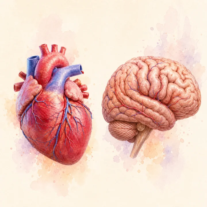
Compare organs
Heart vs. brain
Function animation
Drives double circulation & maintains blood pressure (~120/80 mmHg)
Clinical notes
Common conditions
- Coronary Artery Disease (Atherosclerosis)
- Myocardial Infarction (Heart Attack)
- Hypertension (High Blood Pressure > 140/90 mmHg)
- Cardiac Arrhythmia & Fibrillation
- Heart Valve Stenosis & Regurgitation
- Congestive Heart Failure (CHF)
- Pericarditis
- Congenital Septal Defects (ASD / VSD)
Where it works